

Campero A (1,3); Abarca-Olivas J (2); González-López P (2); Fernández-Cornejo V (2); Verdú-Martinez I (2); Moreno-López P (2); Lloret-García J (2); Rhoton AL Jr (3)
(1) Department of Neurosurgery, Hospital Padilla, Tucumán, Argentina.
(2) Laboratory of Neuroanatomy, Universidad Miguel Hernández de Alicante. Alicante, Spain.
(3) Department of Neurological Surgery, Gainesville, Florida, USA
>Introduction
The posterior petrosectomies involve progressive, stepwise drilling of the petrous bone: The presigmoid retrolabyrinthine, translabyrinthine, and transcochlear approaches are exposed. Transpetrosal approaches progressively flatten the temporal bone to maximize surgical exposure and to minimize retraction on the cerebellum. The surgical corridor between the cerebellum and the petrous bone is also progressively widened during transpetrosal approaches.
Step 0: Position
The patient is placed supine or in the park bench or sitting position. The sitting position has almost been abandoned because it is associated with a significant risk of air embolism. The sitting position, however, does offer a clean field because blood and CSF drain from the surgical field. The head is slightly flexed and rotated toward the ipsilateral side. This position can be uncomfortable for the surgeon, leading to early interruption of the procedure. In the supine position, the patient’s head is rotated toward the contralateral side and is supported on a Mayfield three-point fixation device. A sand bag or a foam roll can be placed under the ipsilateral shoulder to minimize rotation of the neck. This same position is used for all posterior transpetrosal approaches. If the patient’s body habitus precludes contralateral head rotation due to neck compression or an elevated ipsilateral shoulder, a modified park bench position can be used.
After anesthesia is induced, we could place a lumbar drain to help minimize retraction on the cerebellum and to avert a CSF leak after surgery. Frameless stereotactic image guidance and monitoring of the facial nerve and somatosensory evoked potentials are standard adjuncts.
Step 1: Skin and muscle

The skin incision is C-shaped and starts in the temporal region above the zygoma, extends above the ear and downward in the suboccipital area medial to the mastoid process.

The skin flap is reflected forward to the level of the external auditory canal. The temporal and the esternocleidomastoid muscles are exposed.
EAC: External auditory canal; ECM: Esternocleidomastoid muscle;TF: Temporal fascia.
Step 2: Mastoidectomy and Craniotomy
Step 2a: Bone landmarks

The mastoid and the posterior part of the squamosal temporal bone, the lateral part of the squamosal occipital bone and the inferior parietal bone are exposed before performing the craniotomy.
DM: Digastric muscle; EAC: External auditory canal; IOM: inferior oblique muscle; LEM: levator scapulae muscle; MP: mastoid process; SOM: superior oblique muscle.
Step 2b: Mastoidectomy

The cortex over the mastoid bone is removed by using a high-speed drill with a large cutting bur. The anterior border of the bone removal is a slightly curved line that extends between the top of the external auditory meatus and the mastoid tip. The superior limit is a line perpendicular to the first line, extending from the root of the zygoma posteriorly to the asterion: the supramastoid crest. The major landmarks in the mastoid area are the lateral semicircular canal, the mastoid antrum superiorly, and the digastric ridge inferiorly. As the cortical bone is drilled, air cells will begin to be encountered.
MAC: mastoid air cells; SH: spine of Henle; SMC: suprameatal crest.

Bone is drilled 1cm behind the sigmoid sinus, maintaining a uniform depth until the sigmoide sinus is exposed. The sigmoid sinus is skeletonized along its length down through the region of the jugular bulb using a diamond bur. The mastoid air cells are removed anteriorly and superiorly to expose the middle fossa dura. The air cells are drilled further anteriorly to expose the compact bone of the bony labyrinth. Once the semicircular canals are exposed we must know that the facial nerve is situated parallel and 1 to 2 mm anterior to the lateral semicircular canal.
SC: Semicircular canals; SH: Spine of Henle; SS: Sigmoid sinus.

A mastoidectomy has been completed to expose the capsule of the posterior and lateral canals and the tympanic and mastoid facial segments. The mastoid segment of the facial nerve descends through the facial canal and gives rise to the chorda tympani , which passes upward and forward across the tympainc membrane and the malleus neck.
CT: chorda of tympani; I: incus; JB: jugular bulb; LSC: lateral semicircular canal; M: malleus; PSC: posterior semicircular canal; S: staple; SM: stapedial muscle; SSC: superior semicircular canal; VIIms: mastoid segment of VII cranial nerve; VIIts: tympanic segment of VII cranial nerve.
Step 2c: Craniotomy

In the combined supra and infratentorial presigmoide approach a temporooccipital craniotomy is performed and the transverse sinus, the superior petrosal sinus and the sigmoid sinus are exposed.
C: cerebellum; JB: jugular bulb; SPS: superior petrosal sinus; SS: sigmoid sinus; TL: temporal lobe; TS: transverse sinus.
Step 3: Durotomy

Trautman’s triangle is exposed with the black lined area. This is the area of dural openning bounded by the superior petrosal sinus, the sigmoid sinus and the bone labyrinth.
SS: sigmoid sinus; TS: Transverse sinus; SPS: superior petrosal sinus; JB: jugular bulb; Black lined area: Trautman’s triangle. The size of this triangle is highly variable depending on the size of the sigmoid sinus.

The temporal supratentorial dura has beeen removed along the base of the temporal craniotomy while preserving the junction of the vein of Labbé with the transverse sinus.

The dural incision is extended across the superor petrosal sinus to join the dural incision in the temporal dura. After division of the superior petrosal sinus, the tentorium is incised parallel to and just behind the petrosal ridge and superior petrosal sinus. This dural incision is extended from the site of division of the superior petrosal sinus through the medial edge of the tentorium to the incisura behind where the trochlerar nerve enters the tentorial edge. The posterior portion of the temporal lobe is elevated and the sigmoid sinus is displaced posteriorly along with the cerebellar hemisphere while preserving the junction of the vein of Labbé with the sigmoid sinus.
AE: arcuate eminence; AICA: anteroinferior cerebellar artery; JB: jugular bulb; SC: semicircular canals; SCA: superior cerebellar artery; SPV: superior petrosal vein; SS: sigmoid sinus; VA: vertebral artery.
Step 4: Intradural stage
Step 4a: retrolabyrinthine variant

Wide view of the cerebellopontine components and their relationship with the bony labyrinth. In this specimen we have performed a retrolabyrinthine approach.
AICA: anteroinferior cerebellar artery; ES: endolymphatic sac; LSC: lateral semicircular canal; PSC: posterior semicircular canal; SCA: superior cerebellar artery; SPV: superior petrosal vein; SS: sigmoid sinus; SSC: superior semicircular canal; VA: vertebral artery.
Step 4b: translabyrinthine variant

In this case a translabyrinthine approach is performed. The semicircular canals and vestibule are resected resulting in the loss of hearing but providing excelent acces to the internal auditory canal.
IAC: internal acustic canal; MF: middle fossa; SMT: suprameatal tubercle; TE: tentorial edge.

In this view the complete facial nerve is skeletonized from its cisternal to its mastoid segment.
CT: chorda of tympani; I: incus; GG: geniculate ganglion; GPN: greater petrosal nerve; M: malleus; S: stapes; SMT: suprameatal tubercle; TS: transverse crest; VII-mes: meatal segment; VII-ls: labyrinthine segment; VII-ts: tympanic segment; VII-ms: mastoid segment.

Here the facial nerve is transposed and moved posteriorly. Note the wide exposure of the lateral and anterior tentorial incisure.
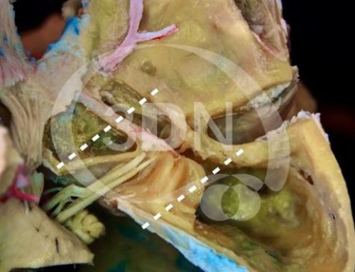
Step 4c: transcochlear approach

The next more extensive modification is the transcochlear approach, in which the cochlea located anteromedial to the fundus of the meatus is removed, thus providing access to the medial part of the petrous apex and the side of the clivus. Another modification, which we call the extended translabyrinthine approach, and is similar to the transcochlear approach, involves drilling bone both anterior and posterior to the facial nerve, leaving the facial nerve skeletonized in a column of bone and working both anterior and posterior to the facial nerve to remove the cochlea and access the side of the clivus. Gaining access for drilling the cochlea anterior to the facial nerve commonly requires that at least part of the posterior part of the external canal be removed, that the tympanic cavity be obliterated, and that the internal carotid artery be exposed below the promontory.
BA: basilar artery; C: cerebellum; MF: middle fosa; P: pons; SPS: superior petrosal sinus; VA: vertebral artery.
References
1-Fossett DT, Caputy AJ. Transpetrosal approach. Felicity Edge, editor. Operative neurosurgical anatomy. 1st ed. New York: Thieme; 2002. p. 67-80.
2-House WF: Middle cranial fossa approach to the petrous pyramid. Arch Otolaryngol 78:460–469, 1963.
3-House WF: Evolution of transtemporal bone removal of acoustic tumors. Arch Otolaryngol 80:731–742, 1964.
4-House WF, Hitselberger WE: The transcochlear approach to the skull base. Arch Otolaryngol 102:334–342, 1976.
5-Rhoton AL Jr; The temporal bone and transtemporal approaches. Neurosurgery, Vol. 47, No. 3, September 2000 Supplement.


Follow us